
Medical imaging
Extensive range of advanced medical imaging services for morphological bone investigations
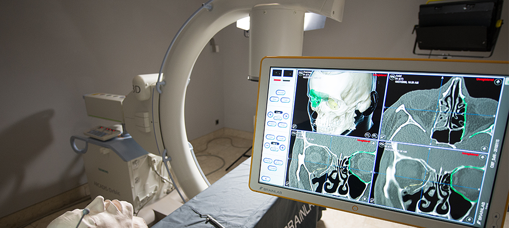
Image acquisition
- Wide range of imaging modalities clinical CT, HR-pQCT, microCT (in vivo/ex vivo), Ultrasound
- External sources: MRI, DXA, Laser Scanning
- In vivo, ex vivo, and in vitro measurement capabilities (ranging from patients to bioresorbable scaffolds)
Image processing
- Data preprocessing (importing, scaling, cropping, aligning)
- Data filtering and segmentation
- 3D bone model generation
Image analysis
- Image based morphological studies
- 3D statistical modeling
- Computerized preoperative planning
- Development of surgical strategies
- Bone density assessment
- Bone microarchitecture evaluation
- Monitoring of fracture healing/bone evolution
- Large bone defects, delayed union models, infection models
- Micro finite element analysis
- Optimization of implant anchorage

l-r: original, segmented, fracture reduction

Qualifications
- Multidisciplinary experienced team (mechanical engineers, physicists, medical and dental doctors, computer scientists)
- Qualified staff for working with experimental animals
- Close collaboration with expert surgeons
Certification
- ISO 9001
- Good Laboratory Practice (GLP)
Contact

Ulrich Bentz
AO Research Institute Davos
Clavadelerstrasse 8
7270 Davos Platz
Switzerland
Tel.: +41 81 414 23 26


